植物幹細胞電顕アトラス
イメージング解析や電子顕微鏡(電顕)技法により植物の幹細胞系譜を追う解析は、これまでほとんど行われていませんでした。新学術領域「植物多能性幹細胞」において当研究室では、最新の蛍光イメージング法と広域2次元および3次元電顕解析法、そして光-電子相関顕微鏡法により幹細胞を特定する技術開発を行うとともに、それら技術を用いて幹細胞の特徴や幹細胞系譜の形成過程の解明を目指し、解析を行いました。
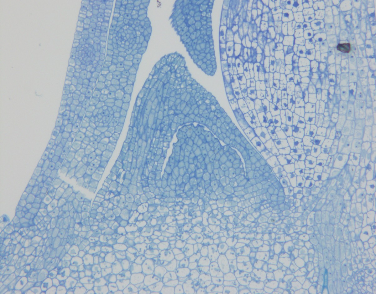
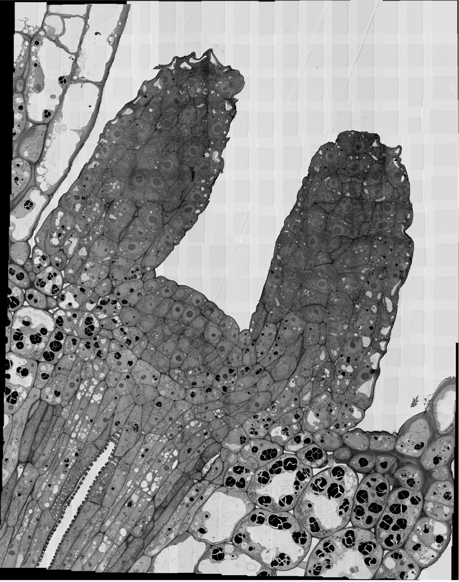
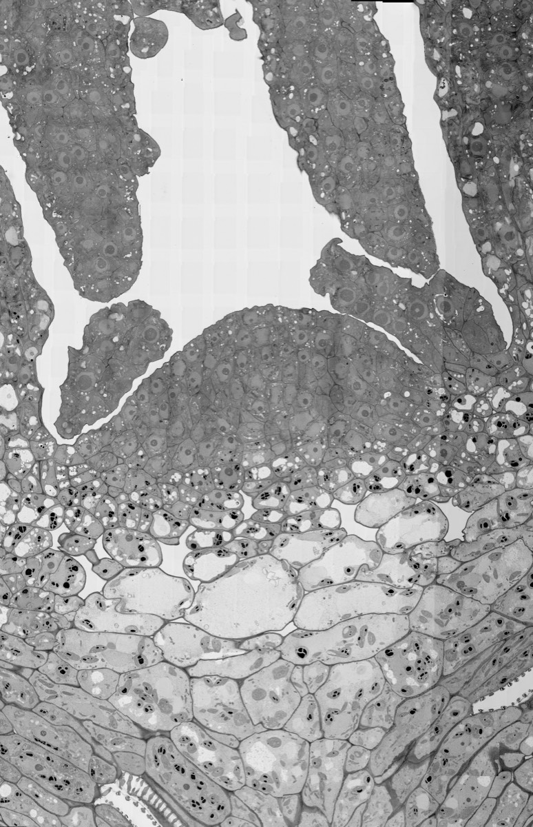
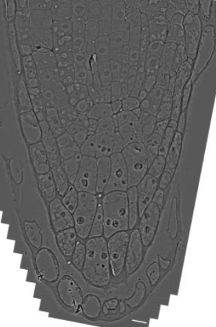
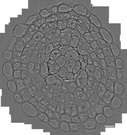
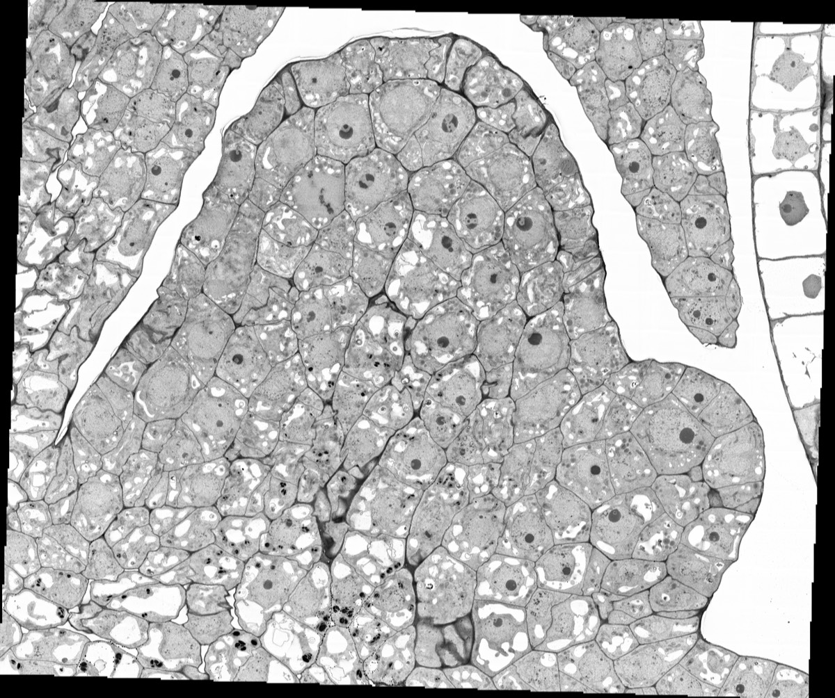
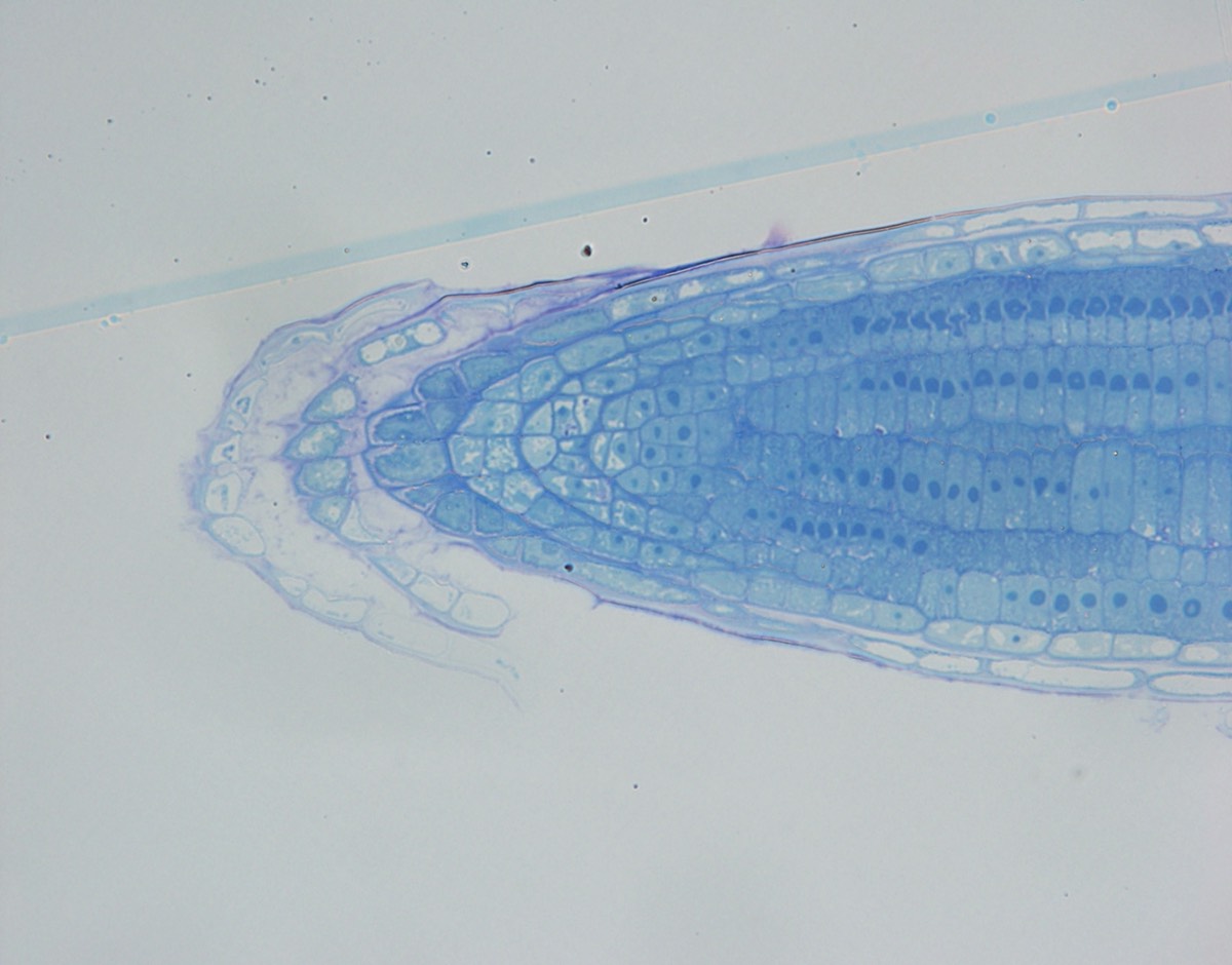
走査電顕(SEM)の高分解能化により、樹脂切片をスライドガラスまたはウェハーなどの基板に載せ、反射電子を検出することで、透過電顕のような画像を撮影する方法 “切片SEM法”が電顕解析の主流に変わりつつあります。この切片SEM法を用いた広域電顕撮像技術により、シロイヌナズナ・イネの茎頂・根端のギガピクセルクラスの高解像度広域電顕写真を取得しました。このような広域電顕写真を多くの研究者や一般の方々に閲覧できるように、Website上で拡大縮小して俯瞰できるGoogle MapsのようなWebsite “電顕アトラス”を構築しています。ここでは、幹細胞電顕アトラス"Plant Stem Cell EM atlas"として、この領域で撮像したシロイヌナズナ・イネの茎頂や根端の広域電顕像を集めました。
To date, few studies have been conducted to trace plant stem cell lineages using imaging and electron microscopy techniques. In the new academic field of "Plant Pluripotent Stem Cells," our laboratory has developed techniques to identify stem cells using state-of-the-art fluorescence imaging, wide-area 2D and 3D electron microscopy, and photoelectron correlation microscopy, and has analyzed stem cell characteristics and stem cell lineage formation processes using these techniques. We used these techniques to analyze the characteristics of stem cells and stem cell lineage formation. With the development of high-resolution scanning electron microscopes (SEM), the "section SEM" method, in which a resin section is placed on a glass slide or a substrate such as a wafer and the reflected electrons are detected, is becoming the mainstream method for electron microscopy analysis. Using the wide-area electron microscopy imaging technique based on the section SEM method, we obtained gigapixel-class high-resolution wide-area electron micrographs of the stem tops and root tips of Arabidopsis thaliana and rice plants. In order to make these wide-area electron microscope images accessible to a large number of researchers and the general public, we have developed a website, "Electron microscope atlas," which is similar to Google Maps and allows users to zoom in and out to get a bird's-eye view of the images. Here, we have collected wide-area electron microscopy images of Arabidopsis and rice stem tops and root tips taken in this area as the "Plant Stem Cell EM atlas.
Arabidopsis
Rice
Slidestacks
Arabidopsis
Rice